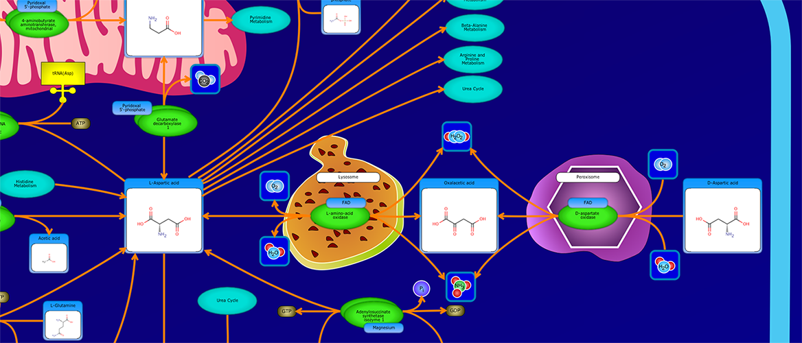
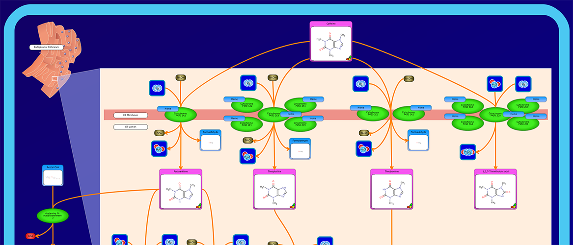
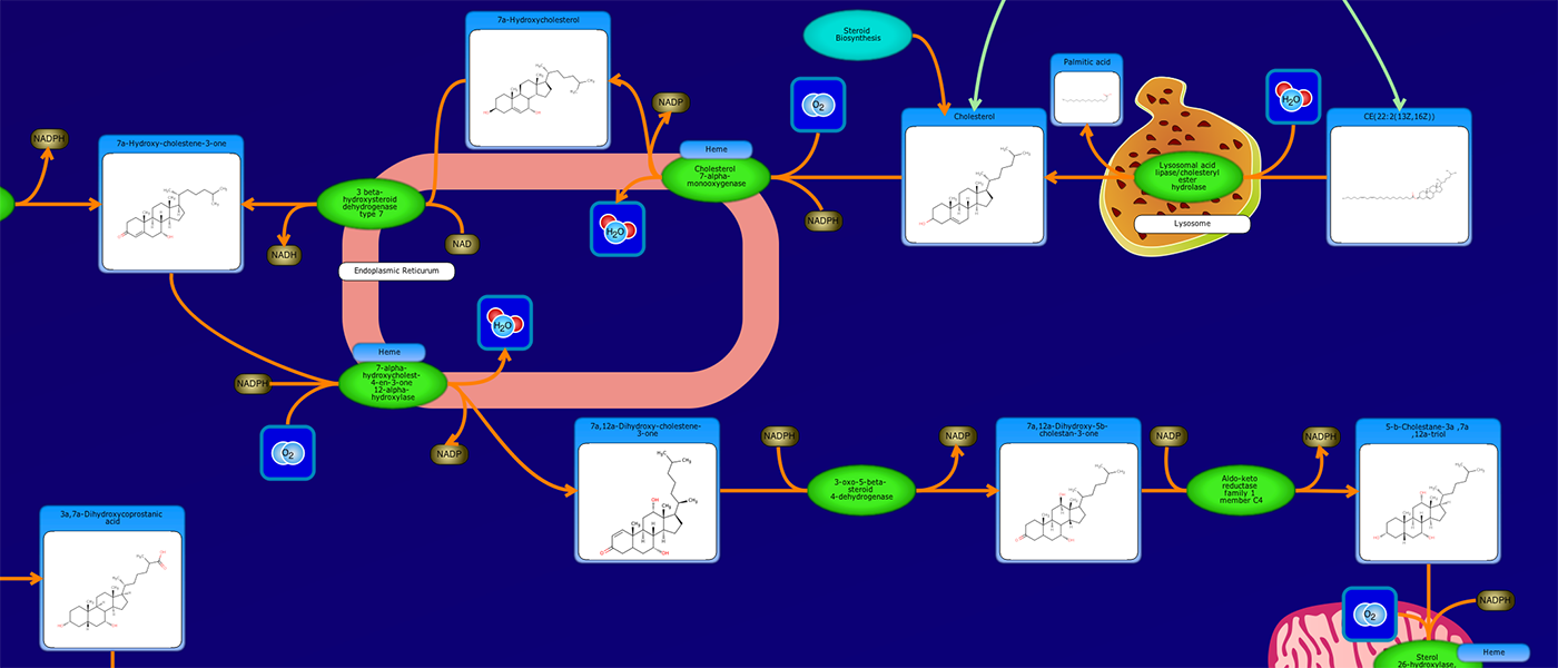
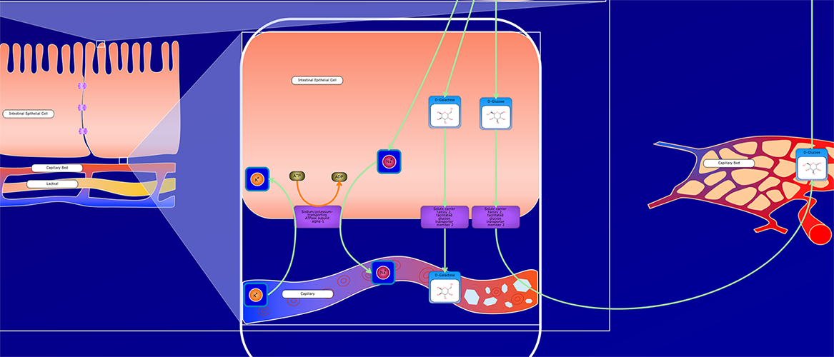
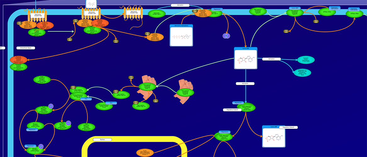
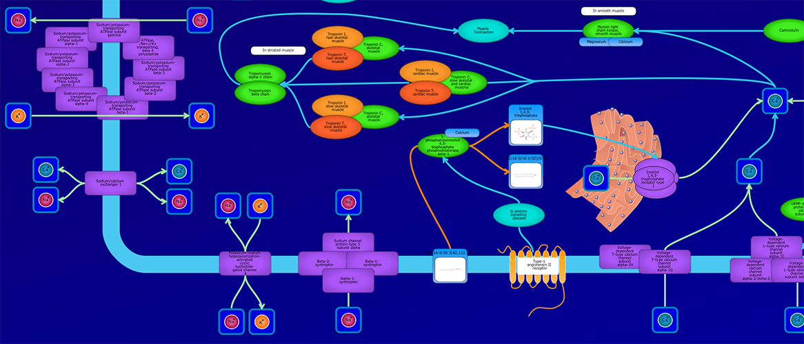
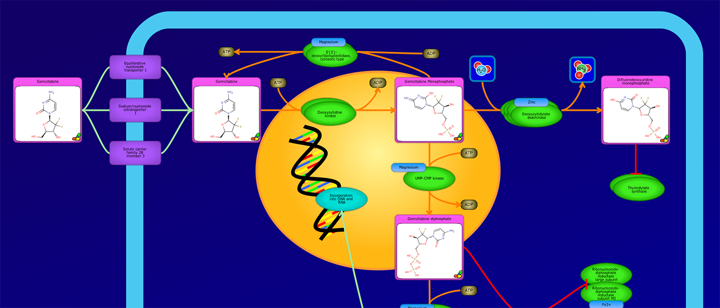
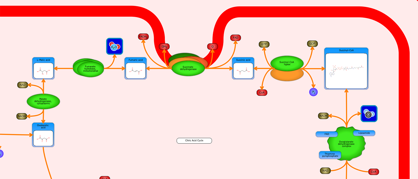
Loading Pathway...
Error: Pathway image not found.
Hide
Pathway Description
Peptidoglycan Biosynthesis I
Escherichia coli
Metabolic Pathway
Peptidoglycan is a net-like polymer which surrounds the cytoplasmic membrane of most bacteria and functions to maintain cell shape and prevent rupture due to the internal turgor. In E. coli K-12, the peptidoglycan consists of glycan strands of alternating subunits of N-acetylglucosamine (GlcNAc) and N-acetylmuramic acid (MurNAc) which are cross-linked by short peptides. The pathway for constructing this net involves two cell compartments: cytoplasm and periplasmic space.
The pathway starts with a beta-D-fructofuranose going through a mannose PTS permease, phosphorylating the compound and producing a beta-D-fructofuranose 6 phosphate. This compound can be obtained from the glycolysis and pyruvate dehydrogenase or from an isomerization reaction of Beta-D-glucose 6-phosphate through a glucose-6-phosphate isomerase. The compound Beta-D-fructofuranose 6 phosphate and L-Glutamine react with a glucosamine fructose-6-phosphate aminotransferase, thus producing a glucosamine 6-phosphate and a l-glutamic acid. The glucosamine 6-phosphate interacts with phosphoglucosamine mutase in a reversible reaction producing glucosamine-1P. Glucosamine-1p and acetyl-CoA undergo acetylation through a bifunctional protein glmU releasing Coa and a hydrogen ion and producing a N-acetyl-glucosamine 1-phosphate. Glmu, being a bifunctional protein catalyzes the interaction of N-acetyl-glucosamine 1-phosphate, hydrogen ion and UTP into UDP-N-acetylglucosamine and pyrophosphate. UDP-N-acetylglucosamine then interacts with phosphoenolpyruvic acid and a UDP-N acetylglucosamine 1- carboxyvinyltransferase releasing a phosphate and the compound UDP-N-acetyl-alpha-D-glucosamine-enolpyruvate.
The latter undergoes a NADPH dependent reduction producing a UDP-N-acetyl-alpha-D-muramate catalyzed by a UDP-N-acetylenolpyruvoylglucosamine reductase. UDP-N-acetyl-alpha-D-muramate and L-alanine react in an ATP-mediated ligation through a UDP-N-acetylmuramate-alanine ligase releasing an ADP, hydrogen ion, phosphate and a UDP-N-acetylmuramoyl-L-alanine. Next, UDP-N-acetylmuramoylalanine-D-glutamate ligase catalyzes an ATP, D-glutamic acid and UDP-N-acetylmuramoyl-L-alanine releasing ADP, phosphate and UDP-N-acetylmuramoyl-L-alanyl-D-glutamate. The latter product then interacts with meso-diaminopimelate in an ATP mediated ligation through a UDP-N-acetylmuramoylalanine-D-glutamate-2,6-diaminopimelate ligase resulting in ADP, phosphate, hydrogen ion and UDP-N-Acetylmuramoyl-L-alanyl-D-gamma-glutamyl-meso-2,6-diaminopimelate. This compound in turn with D-alanyl-D-alanine react in an ATP-mediated ligation through UDP-N-Acetylmuramoyl-tripeptide-D-alanyl-D-alanine ligase to produce UDP-N-acetyl-alpha-D-muramoyl-L-alanyl-gama-D-glutamyl-meso-2,6-diaminopimeloyl-Dalanyl-D-alanine and hydrogen ion, ADP, phosphate. UDP-N-acetyl-alpha-D-muramoyl-L-alanyl-gama-D-glutamyl-meso-2,6-diaminopimeloyl-Dalanyl-D-alanine interacts with di-trans,octa-cis-undecaprenyl phosphate through a phospho-N-acetylmuramoyl-pentapeptide-transferase, resulting in UMP and Undecaprenyl-diphospho-N-acetylmuramoyl-L-alanyl-D-glutamyl-meso-2,6-diaminopimeloyl-D-alanyl-D-alanine which in turn reacts with a UDP-N-acetylglucosamine through a N-acetylglucosaminyl transferase to produce a hydrogen, UDP and ditrans,octacis-undecaprenyldiphospho-N-acetyl-(N-acetylglucosaminyl)muramoyl-L-alanyl-gamma-D-glutamyl-meso-2,6-diaminopimeloyl-D-alanyl-D-alanine. This compound ends the cytoplasmic part of the pathway. ditrans,octacis-undecaprenyldiphospho-N-acetyl-(N-acetylglucosaminyl)muramoyl-L-alanyl-gamma-D-glutamyl-meso-2,6-diaminopimeloyl-D-alanyl-D-alanine is transported through a lipi II flippase. Once in the periplasmic space, the compound reacts with a penicillin binding protein 1A prodducing a peptidoglycan dimer, a hydrogen ion, and UDP. The peptidoglycan dimer then reacts with a penicillin binding protein 1B producing a peptidoglycan with D,D, cross-links and a D-alanine.
References
Peptidoglycan Biosynthesis I References
Bouhss A, Trunkfield AE, Bugg TD, Mengin-Lecreulx D: The biosynthesis of peptidoglycan lipid-linked intermediates. FEMS Microbiol Rev. 2008 Mar;32(2):208-33. doi: 10.1111/j.1574-6976.2007.00089.x. Epub 2007 Dec 10.
Pubmed: 18081839
Bupp K, van Heijenoort J: The final step of peptidoglycan subunit assembly in Escherichia coli occurs in the cytoplasm. J Bacteriol. 1993 Mar;175(6):1841-3.
Pubmed: 8449890
Cooper S: Synthesis of the cell surface during the division cycle of rod-shaped, gram-negative bacteria. Microbiol Rev. 1991 Dec;55(4):649-74.
Pubmed: 1779930
Ikeda M, Wachi M, Jung HK, Ishino F, Matsuhashi M: The Escherichia coli mraY gene encoding UDP-N-acetylmuramoyl-pentapeptide: undecaprenyl-phosphate phospho-N-acetylmuramoyl-pentapeptide transferase. J Bacteriol. 1991 Feb;173(3):1021-6.
Pubmed: 1846850
Inoue A, Murata Y, Takahashi H, Tsuji N, Fujisaki S, Kato J: Involvement of an essential gene, mviN, in murein synthesis in Escherichia coli. J Bacteriol. 2008 Nov;190(21):7298-301. doi: 10.1128/JB.00551-08. Epub 2008 Aug 15.
Pubmed: 18708495
Mengin-Lecreulx D, Texier L, Rousseau M, van Heijenoort J: The murG gene of Escherichia coli codes for the UDP-N-acetylglucosamine: N-acetylmuramyl-(pentapeptide) pyrophosphoryl-undecaprenol N-acetylglucosamine transferase involved in the membrane steps of peptidoglycan synthesis. J Bacteriol. 1991 Aug;173(15):4625-36.
Pubmed: 1649817
Mohammadi T, van Dam V, Sijbrandi R, Vernet T, Zapun A, Bouhss A, Diepeveen-de Bruin M, Nguyen-Disteche M, de Kruijff B, Breukink E: Identification of FtsW as a transporter of lipid-linked cell wall precursors across the membrane. EMBO J. 2011 Apr 20;30(8):1425-32. doi: 10.1038/emboj.2011.61. Epub 2011 Mar 8.
Pubmed: 21386816
Mohammadi T, Sijbrandi R, Lutters M, Verheul J, Martin NI, den Blaauwen T, de Kruijff B, Breukink E: Specificity of the transport of lipid II by FtsW in Escherichia coli. J Biol Chem. 2014 May 23;289(21):14707-18. doi: 10.1074/jbc.M114.557371. Epub 2014 Apr 7.
Pubmed: 24711460
Ruiz N: Bioinformatics identification of MurJ (MviN) as the peptidoglycan lipid II flippase in Escherichia coli. Proc Natl Acad Sci U S A. 2008 Oct 7;105(40):15553-7. doi: 10.1073/pnas.0808352105. Epub 2008 Oct 1.
Pubmed: 18832143
Schleifer KH, Kandler O: Peptidoglycan types of bacterial cell walls and their taxonomic implications. Bacteriol Rev. 1972 Dec;36(4):407-77.
Pubmed: 4568761
Sham LT, Butler EK, Lebar MD, Kahne D, Bernhardt TG, Ruiz N: Bacterial cell wall. MurJ is the flippase of lipid-linked precursors for peptidoglycan biogenesis. Science. 2014 Jul 11;345(6193):220-2. doi: 10.1126/science.1254522.
Pubmed: 25013077
Van Heijenoort J, Elbaz L, Dezelee P, Petit JF, Bricas E, Ghuysen JM: Structure of the meso-diaminopimelic acid containing peptidoglycans in Escherichia coli B and Bacillus megaterium KM. Biochemistry. 1969 Jan;8(1):207-13.
Pubmed: 4975910
van Heijenoort J: Formation of the glycan chains in the synthesis of bacterial peptidoglycan. Glycobiology. 2001 Mar;11(3):25R-36R.
Pubmed: 11320055
WEIDEL W, PELZER H: BAGSHAPED MACROMOLECULES--A NEW OUTLOOK ON BACTERIAL CELL WALLS. Adv Enzymol Relat Subj Biochem. 1964;26:193-232.
Pubmed: 14150645
Highlighted elements will appear in red.
Highlight Compounds
Highlight Proteins
Enter relative concentration values (without units). Elements will be highlighted in a color gradient where red = lowest concentration and green = highest concentration. For the best results, view the pathway in Black and White.
Visualize Compound Data
Visualize Protein Data
Downloads
Settings